Friday, February 19, 2010

Cervical Cancer


Many women pass symptoms of cervical cancer off as PMS. Often by the time symptoms accumulate the cancer is far progressed. Symptoms of cervical cancer include: unusual heavy discharge, pelvic pain that is not related to the menstrual cycle, dysuria, bleeding between cycles or after intercourse or a pelvic exam.

Increased risk factors include having multiple sexual partners or a partner with multiple sexual partners, having many children, being on birth control pills for extended time - however studies have shown that a woman's risk is dramatically reduced after ceasing taking them and a lack of pap smears

Once an abnormal pap smear result is received a biopsy will be performed to give a more definitive diagnosis. Once cervical cancer is found, CT and MRI's are often done to ensure the cancer has not spread, causing metastatic disease. A PET scan could be performed to stage the cancer and help plan the best radiation or chemotherapy to fight the cancer after surgery.

Cervical cancer is one of the most preventable types of cancers. With regular pap smears cervical cancer can be prevented in most cases. Survival rates have gone up tremendously since more and more women get an annual pap smear.

Saturday, February 6, 2010

Pancreatitis


Acute pancreatitis is an inflammation of the pancreas. The pancreas is an organ that secretes insulin and glucagon and also aids in digestion.


Symptoms most often include left upper quadrant abdominal pain, fever, nausea and vomiting. This disease is often is a result of gallbladder dysfunction or alcoholism.


Treatment for pancreatitis usually includes IV pain medication due to the severe abdominal pain. It also requires IV fluids and withholding food to attempt to keep the pancreas inactive. This allows time for the inflamed pancreas to resolve. If medication does not work, then the next steps include removing gallstones or to place a stent in the pancreas and to drain fluid from around the pancreas. This is done oftentimes in Radiology under fluoro during a procedure called ERCP or endoscopic retrograde cholangiopancreatography.


This disease is treatable and nearly all recover. However, there is a small percentage that can develop chronic pancreatitis as well.

Wednesday, January 27, 2010

Empyema


Empyema is a collection of pus in between the space of the lung and chest wall. It is an infection that spreads from the lungs and leads to a build up of fluid in the pleural space. Symptoms of this condition include chest pain, cough, dyspnea, fever, and sometimes weight loss.
Empyema has several possible causes but is most often is an associated complication of pneumonia.


There are three phases of empyema. The acute phase that fills the lung cavity with thin purulent fluid, second stage in which the fluid thickens, and then a third stage in which the lung is covered with a thick fibrous material. The third phase can only be cured with a thoracotomy to remove that thick fluid and scrape the lining of the lung of the fibrous material. In the earlier phases antibiotics or drainages can help clear the infection.

When this infection complicates pneumonia it raises the risk of death and permanent damage to lung tissue significantly. But, with long term antibiotic therapy and drainages the patient most often recovers.

Tuesday, December 1, 2009

Kyphoplasty for compression fractures of thoracic spine


When particular patients are assessed after a spinal fracture and meet certain guidelines, their physician may recommend a procedure called kyphoplasty. These patients usually have a pathologic fracture or a fracture due to osteoporosis. Osteoporosis weakens the bone and makes the simplest fall or cough a major problem.

The goal of this procedure is to help relieve pain from the spine due to fracture and to stabilize the spine as well. The other goal is to return loss of height to the vertebral body.

The exam is usually done in a Special Procedures lab or an OR setting. The physician will insert a balloon into the vertebrae and inject a special filler that is a hard cement like material that expands. This assists with the bone height loss due to the compression fracture. After the procedure is over many patients immediately feel relief. To document the placement a CT scan is done post procedure. I've attached some images of a pre kyphoplasty patient with a t12 compression fracture and then the same patient's post exam.

Burst Fractures


Burst fractures most often occur in the thoracolumbar spine, around T-11 through L2. Burst fractures are usually the result of some sort of trauma to the spine such as a motor vehicle accident or fall. These fractures can occur in patients of any age. Treatment does vary for these injuries. Neurosurgeons will evaluate images from CT and MRI. If the patient is not paralyzed and has no significant canal compromise then normally a brace is in order for the patient. The physician will monitor healing by obtaining lateral radiographs of the fracture sight to document healing. If patient has paralysis that is not resolving with canal compromise, then surgery to decompress the stenosis will need to be performed. I have attached some CT images demonstrating this deformity.

Friday, November 20, 2009

Cervical disc bulge


We received a 61 y/o male through the emergency room for a CT cervical spine among everything else ordered. He was alert and orientated and was responding to questions. However, he had tingling sensation to the nipple line, but was unable to feel anything from that point down. He suffered a 4 foot fall from a truck bed.


The exam was performed and it was noted that he had a significant C-3 fracture with significant offset. All other imaging studies were negative. After a short time in the emergency room, the patient's respiratory effort declined significantly and had to be intubated.


The following day a CT cervical myelogram was ordered. An attempt by the Radiologist was made, but due to equipment and patient condition a blind stick by the Neurosurgeon in the ICU was performed. The patient then returned to the CT suite to have post images performed.

I've attached the reformats performed and it shows a moderate to severe disc bulge. This certainly can be causing his paralysis.

Neurosurgeon re-evaluated patient and states he is ineligible for MRI due to pain stimulator implant. He will give the patient another day to recover and see him again to evaluate stability with flexion and extension movements. He also states that he has cord contusion that can resolve with some time.

Due to the numbness/tingling and inability to feel past that point the ED physician felt certain that there was a spine injury. These injuries can resolve on their own or often require surgery to assist healing.

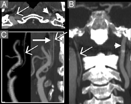
Carotid Stenosis and Strokes


Carotid stenosis is a narrowing of the artery due to a build up of plaque. The carotid arteries are what feeds the brain. When there is a major stenosis or plaque build up it can cause an ischemic stroke, the most common stroke diagnosed.


Stroke symptoms range from weakness in extremities, slurred speech, headache, facial droop, and trouble with gait among others. To evaluate a patient for a stroke, normally a routine CT head is done to rule out a bleed. Once this is negative, a carotid ultrasound may be ordered. Oftentimes now, a CTA head and neck is ordered from the ER. When a CTA is performed it is specifically looking at the vessels. Stenosis can be determined with precision accuracy rather than traditional angiography which is more invasive and carries some additional risks.


Treatment for this condition depends on the results but ranges from medication to surgery. The most important rule for stroke is early intervention.
The picture at the left top shows an ICA with near total occlusion.

The picture at left bottom shows carotid stenosis which is the dark spot by the area.