Friday, November 20, 2009

Cervical disc bulge


We received a 61 y/o male through the emergency room for a CT cervical spine among everything else ordered. He was alert and orientated and was responding to questions. However, he had tingling sensation to the nipple line, but was unable to feel anything from that point down. He suffered a 4 foot fall from a truck bed.


The exam was performed and it was noted that he had a significant C-3 fracture with significant offset. All other imaging studies were negative. After a short time in the emergency room, the patient's respiratory effort declined significantly and had to be intubated.


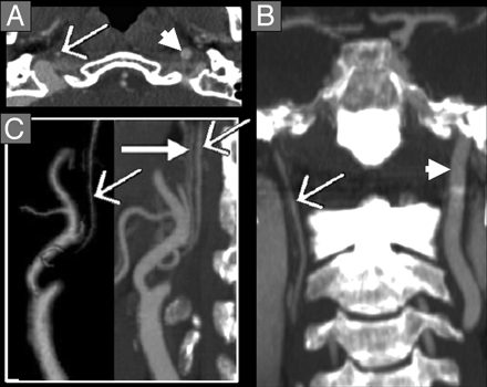
The following day a CT cervical myelogram was ordered. An attempt by the Radiologist was made, but due to equipment and patient condition a blind stick by the Neurosurgeon in the ICU was performed. The patient then returned to the CT suite to have post images performed.

I've attached the reformats performed and it shows a moderate to severe disc bulge. This certainly can be causing his paralysis.

Neurosurgeon re-evaluated patient and states he is ineligible for MRI due to pain stimulator implant. He will give the patient another day to recover and see him again to evaluate stability with flexion and extension movements. He also states that he has cord contusion that can resolve with some time.

Due to the numbness/tingling and inability to feel past that point the ED physician felt certain that there was a spine injury. These injuries can resolve on their own or often require surgery to assist healing.

Carotid Stenosis and Strokes


Carotid stenosis is a narrowing of the artery due to a build up of plaque. The carotid arteries are what feeds the brain. When there is a major stenosis or plaque build up it can cause an ischemic stroke, the most common stroke diagnosed.


Stroke symptoms range from weakness in extremities, slurred speech, headache, facial droop, and trouble with gait among others. To evaluate a patient for a stroke, normally a routine CT head is done to rule out a bleed. Once this is negative, a carotid ultrasound may be ordered. Oftentimes now, a CTA head and neck is ordered from the ER. When a CTA is performed it is specifically looking at the vessels. Stenosis can be determined with precision accuracy rather than traditional angiography which is more invasive and carries some additional risks.


Treatment for this condition depends on the results but ranges from medication to surgery. The most important rule for stroke is early intervention.
The picture at the left top shows an ICA with near total occlusion.

The picture at left bottom shows carotid stenosis which is the dark spot by the area.


Saturday, November 14, 2009

Malignant Parotid Tumor


Malignant tumors of the parotid gland are actually quite rare. They occur at a rate of 1-2 cases per 100,000. These tumors occur in slightly more women than men and also have a higher rate of incidence in the Eskimo population. Mortality will depend on the characteristic of the tumor and the stage of the tumor. Some tumors are slow growing and just need to be watched and others are very agressive and require surgery. Sometimes these tumors can grow back. Pain is not necessarily an indicator that this tumor is malignant, but it can be an indicator of progression in a patient diagnosed with a malignancy.


Most tumors affect adults aged 30-70, but vary in stages and malignancy. Tumors that do occur in children have a much higher incidence of malignancy, around 35%.


CT and MRI are both imaging modalities of choice for these tumors for different reasons. CT will best demonstrate the inflammatory, recurrent mass and MRI is best for those masses that are not painful. These tests combined with a fine needle aspiration to determine cellular structure will diagnose this condition with near precision accuracy.