Friday, November 20, 2009

Cervical disc bulge


We received a 61 y/o male through the emergency room for a CT cervical spine among everything else ordered. He was alert and orientated and was responding to questions. However, he had tingling sensation to the nipple line, but was unable to feel anything from that point down. He suffered a 4 foot fall from a truck bed.


The exam was performed and it was noted that he had a significant C-3 fracture with significant offset. All other imaging studies were negative. After a short time in the emergency room, the patient's respiratory effort declined significantly and had to be intubated.


The following day a CT cervical myelogram was ordered. An attempt by the Radiologist was made, but due to equipment and patient condition a blind stick by the Neurosurgeon in the ICU was performed. The patient then returned to the CT suite to have post images performed.

I've attached the reformats performed and it shows a moderate to severe disc bulge. This certainly can be causing his paralysis.

Neurosurgeon re-evaluated patient and states he is ineligible for MRI due to pain stimulator implant. He will give the patient another day to recover and see him again to evaluate stability with flexion and extension movements. He also states that he has cord contusion that can resolve with some time.

Due to the numbness/tingling and inability to feel past that point the ED physician felt certain that there was a spine injury. These injuries can resolve on their own or often require surgery to assist healing.

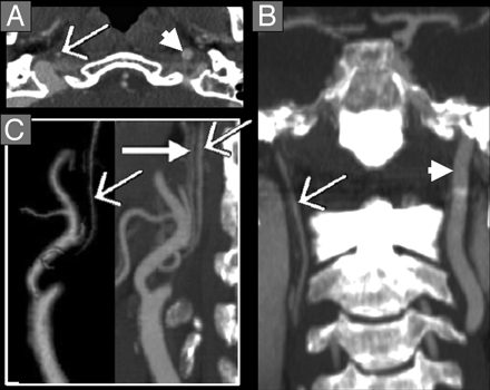
Carotid Stenosis and Strokes


Carotid stenosis is a narrowing of the artery due to a build up of plaque. The carotid arteries are what feeds the brain. When there is a major stenosis or plaque build up it can cause an ischemic stroke, the most common stroke diagnosed.


Stroke symptoms range from weakness in extremities, slurred speech, headache, facial droop, and trouble with gait among others. To evaluate a patient for a stroke, normally a routine CT head is done to rule out a bleed. Once this is negative, a carotid ultrasound may be ordered. Oftentimes now, a CTA head and neck is ordered from the ER. When a CTA is performed it is specifically looking at the vessels. Stenosis can be determined with precision accuracy rather than traditional angiography which is more invasive and carries some additional risks.


Treatment for this condition depends on the results but ranges from medication to surgery. The most important rule for stroke is early intervention.
The picture at the left top shows an ICA with near total occlusion.

The picture at left bottom shows carotid stenosis which is the dark spot by the area.


Saturday, November 14, 2009

Malignant Parotid Tumor


Malignant tumors of the parotid gland are actually quite rare. They occur at a rate of 1-2 cases per 100,000. These tumors occur in slightly more women than men and also have a higher rate of incidence in the Eskimo population. Mortality will depend on the characteristic of the tumor and the stage of the tumor. Some tumors are slow growing and just need to be watched and others are very agressive and require surgery. Sometimes these tumors can grow back. Pain is not necessarily an indicator that this tumor is malignant, but it can be an indicator of progression in a patient diagnosed with a malignancy.


Most tumors affect adults aged 30-70, but vary in stages and malignancy. Tumors that do occur in children have a much higher incidence of malignancy, around 35%.


CT and MRI are both imaging modalities of choice for these tumors for different reasons. CT will best demonstrate the inflammatory, recurrent mass and MRI is best for those masses that are not painful. These tests combined with a fine needle aspiration to determine cellular structure will diagnose this condition with near precision accuracy.


Saturday, October 31, 2009

CTA versus conventional angiogram or DSA in diagnosing aneurysms


The picture at the left is a right middle cerebral saccular aneurysm demonstrated on an MRA. The above picture is a great comparison of a CTA image of a rt posterior inferior cerebral aneurysm and then the same on a DSA image. The DSA image is the conventional angiographic approach to diagnosis. CTA, computed tomography angiography, has nearly replaced traditional angiography in many cases. CTA is faster and non-invasive, whereas angiography carries more risk. There is a femoral stick and often sedation for the patient. More cost is involved as well. A physician must perform this exam. It is also a slower process with some recovery time involved. Physicians most often are turning to CTA first as a guide to finding the answers they need. This has not removed the need for MRA, however. Most neurosurgeons require both a CTA and a MRA pre-op to ensure all their bases are covered.
There was a study published in the Open Neurology Journal in January 2009 regarding the efficacy of CTA vs traditional approaches. They looked at how effective CTA was in pre-operative planning. They found that 72% of the cases CTA was the best imaging quality to define the aneurysm. There were no cases that DSA demonstrated aneurysms that CTA simply missed. They only significant reason for performing the DSA was simply for blood flow demonstration.
I think this study definitely makes the case for CTA and I think with the advent of more platforms such as Vitrea and Tera Recon and upgrades, our reformats will just get better.
Susan Brumley

Friday, October 9, 2009

Temporal bone fractures


Temporal bone fractures occur in the transverse plane and longitudinal plane. These fractures are the result of blunt force trauma. They can result in hearing loss, visual disturbances, facial paralysis, and vertigo. Sometimes these symptoms may be delayed.


Prior to CT's improved detail and spatial resolution, imaging didn't play a big role in diagnosing this. Now with the advent of multi-slice and 3 D reconstruction, CT is at the fore front of this treatment plan. Most patients will present quickly as they will also have basilar skull fractures or raccoon eyes. If a temporal bone fracture goes undiagnosed initially, the patient may present with CSF otorrhea or hearing dysfunction.


Most of these type fractures resolve on their own. Usually, these patients present with much more serious injuries ranging from whole organ injury to skull fractures. These injuries are taken care of first. However, in most cases, even the CSF leak, this resolves in times ranging from 7-14 days to 2-3 weeks for more serious cases.


Retinoblastoma


Retinoblastomas are tumors that occur in the eye. This cancer is one of four of the most common cancers that affect children. The first sign of this disease is usually noted by the white area in the child's eye or eyes. This syndrome can be bilateral. Other symptoms can include red and painful eye or eyes and misaligned eyes. Often, this is hereditary. The average age of diagnosis is 18 months old. Calcifications often occur and that is how this image shows the tumor.


Treatment for this disorder will often involve many specialists including a pediatric oncologist, pediatric opthamologist, and radiation therapist. Most often chemotherapy is given to try to shrink the tumor. Laser therapy, cryotherapy, and external beam irradiation therapy are often utilized to treat this condition. Biopsies are never performed as this can spread metastases to other parts of the body.


This condition is still rather rare with 1 case out of approximately 20,000 live births. If detected early the survival rate is 96% according to the website http://www.retinoblastoma.ca/ Blindness is still a possibility in the affected eye due to the tumor and then treatments to cure the cancer. However, many children only have unilateral retinoblastoma leaving the unaffected eye to do the work.
Susan Brumley


Saturday, September 26, 2009

Cushing's Disease


Cushing’s disease is an endocrine disease that occurs when too much cortisol is in the blood due to excessive ACTH, adrenocorticotrophich hormone. The pituitary is responsible for this anomaly. ACTH stimulates the adrenal glands to secrete cortisol. This disease is most common in those aged 20-70 and is most likely to be women.
Cushing’s disease is not the same as Cushing Syndrome. Cushing’s disease is a direct cause of a pituitary tumor, most likely a pituitary adenoma. Cushing's syndrome mostly means that there is an increased cortisol level, however a pituitary tumor is not the cause. There are many reasons that the body can secrete too much cortisol. Common symptoms of Cushing's disease include skin changes/bruising, weight gain including added fat on the back of the neck, decreased fertility and sex drive, high blood pressure, diabetes, and mood or behavior disorders.
Early signs of Cushing’s disease often go unnoticed because they come on slowly. The first thing most physicians do to determine Cushing’s disease is to do a blood test to determine elevated cortisol in the blood stream. Once Cushing’s Disease is suspected, an MRI is performed. MRI is about 70% accurate in diagnosing this disease.
The only cure for this disorder is transsphenoidal surgery to remove the tumor. If the tumor has not spread to other structures, the cure rate is about 80-85%. Medication is an alternative to surgery. They can be used to help inhibit ACTH production.